Friday 22 August 2014

The ALS Ice Bucket Challenge. What is ALS?

The ALS ice bucket challenge has gone viral on social media. It is an activity involving dumping a bucket of ice water on one's head to create awareness of Amyotrophic lateral sclerosis (ALS) or better known as Lou Gehrig’s Disease.

Amyotrophic lateral sclerosis (ALS), often referred to as "Lou Gehrig's Disease," is a progressive neurodegenerative disease that affects nerve cells in the brain and the spinal cord. Motor neurons reach from the brain to the spinal cord and from the spinal cord to the muscles throughout the body. The progressive degeneration of the motor neurons in ALS eventually leads to their death. When the motor neurons die, the ability of the brain to initiate and control muscle movement is lost. With voluntary muscle action progressively affected, patients in the later stages of the disease may become totally paralyzed.

The challenge dares nominated participants to be filmed having a bucket of ice water poured on their heads with the message spreading the awareness on ALS and make donation to the ALS Association. The person can then send out challenges to other people who must comply within 24 hours. The second form of the challenge requires the participant to donate US$10 to ALS research if they pour the ice water over their head or US$100 if they do not. Good sports do both – then pass on the challenge to someone else.

Among the popular participant that participates in this Ice Bucket Challenge is, Bill Gates, LeBron James, Mark Zuckerberg, Lisa Surihani, Justin Timberlake, and even Malaysian Minister, Khairi Jamaludin. 

Here is one of the video compilation on the Ice Bucket Challenge: 

Find out more about ALS and show your support by donating here www.alsa.org

#icebucketchallenge #alsicebucketchallenge #strikeoutals.

Wednesday 9 July 2014

iGene Second Quarter Newsletter | 2014


As we are now at the third quarter of year 2014, it is a perfect time to look back at the second quarter of 2014 on our work, effort, progression and achievements.

The future belongs to those who believe in the beauty of their dreams.” ― Eleanor Roosevelt

Attached here is iGene second quarter newsletter:
 iGene Second Quarter Newsletter | 2014

Tuesday 8 July 2014

What Comes After Death?


Approximately 105 people die every minute and 2 people die every second,
But the question is, what comes after death? and what can be done by the family to give the last respect to their loved ones?
The answer is in this video, click to watch:


Tuesday 17 June 2014

What Malaysian Says About DIGITAL AUTOPSY?

A public survey was conducted during Festival Belia Putrajaya 2014, to find out how much Malaysian knows about Digital Autopsy (DA).
Two simple question was given which is :
 1. Do you know about Digital Autopsy?
 2. Which one is the best for the community, Classical Autopsy or Digital Autopsy?

Find out more here CLICK ON THE IMAGE BELOW:


ENJOY WATCHING and Please leave your comment on the video by answering those two question.
THANKS

Friday 6 June 2014

UK's second Digital Autopsy facility opens in Bradford, offering non-invasive post-mortem examinations.


State-Of-The-Art Digital Autopsy Facility opens in Bradford on May 30th 2014.
You can find some of the photos here on  FACEBOOK


Friday 2 May 2014

INFOVALLEY First Quater Newsletter | 2014

As we are now at the second quarter of year 2014, it is a perfect time to look back at the first quarter of 2014 on our work, effort, progression and achievements.

"Our life is made up of time. Our days are measured in hours, our pay measured by those hours, our knowledge is measured by years. And yet time eventually runs out and you wonder in your heart of heart if those seconds, minuts, hours, days, weeks, months, years and decades were being spent the best way they possibly could." - Cecelia Ahern

Attached here is INFOVALLEY® Group of Companies first quarter newsletter:

Wednesday 5 February 2014

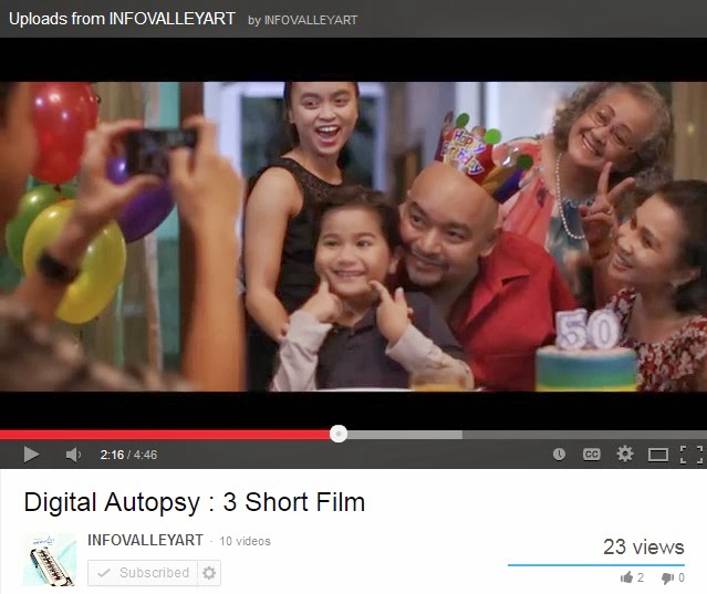
Digital Autopsy : 3 Short Film

The Story of Life, because We Care.

http://www.youtube.com/watch?v=e_wiGXmFREA
Enjoy the 3 short film on Digital Autopsy (Click on the image)


        Digital Autopsy allows expert or forensic pathologist to do autopsy not just once, but they can redo and ask for second opinion. The raw data acquired from the CT modality make it possible as the exact raw data can’t be modified or disturbed. Data can be kept in database with implementation of security measure to be available at any time. This can be kept forever as an archive or for purpose of medical study and research. 

    Digital Autopsy becomes a complement workflow in autopsy procedure. Forensic Pathologist should practice digital autopsy which can aid them to come out with certain finding in the post mortem. With availability of crime scene data, photograph and reconstruction model, Forensic Expert can simulate and test the hypothesis for the manner of death of that particular case. Last but not least, Digital Autopsy can reduce the autopsy time session and give more descriptive and objective visual report. 

Thursday 16 January 2014

Launch of Digital Autopsy Facility in Sheffield

           iGene® Sdn. Bhd has successfully launched the world’s first Digital Autopsy Facility (DAF) network in Sheffield, United Kingdom. The £3million facility, first of the 18 state-of-the-art DAFs was launched on Wednesday 27th November 2013 by Chief Coroner HHJ Peter Thornton QC and Sheffield City Council leader Julie Dore. This new facility in Sheffield, which is housed at the city's Medico-Legal Centre, is intended to pave the way for a series of DAFs nationwide which will cost iGene an approximate £50m.

        The news about the launch has been covered extensively in global media through various mediums including television, radio, and online coverage. The news of the launch has been covered in BBC News, Sky News, Bernama, Google, Daily Mail to name a few.
Below are some of the snapshots from selected news coverage:

BBC Look North News (Click on the image)

Al-Jazeera (Click on the image)
                              

Sky News TV
BT News (Click on the image)

                           
                 The Star UK (Click on the image)               TV3- Buletin Utama                                            The Star UK (Click on the image)

Wednesday 15 January 2014

CSI and Digital Autopsy (Awareness Video)

CSI and Digital Autopsy Animation Video (Click on the image)
The term AUTOPSY has already been adapted and is familiar in the world of Forensic Medicine. The CSI affect more or less influences in appreciative of common people in the field of Forensics Application. Autopsy, also known as post-mortem examination is usually performed by specialized medical doctor in order to determine the cause and manner of death. It normally includes surgery and invasive examination to the dead body. It can be done for either legal or medical purposes. 
DIGITAL AUTOPSY is a complementary approach to classical autopsy. Instead of cutting the cadaver, the dead body will be sent for scanning first, followed by rendering of the body DICOM data into 3D digital body. Forensic expert can perform digital autopsy on the body using the advanced medical visualization software system. If the result is found to be inconclusive, the body can undergoes autopsy in more targeted and educated manner. Digital Autopsy analyzes the 3D volume data generated from Medical Modern Imaging Modalities such as CT scanner or MRI Scanner with direct volume rendering techniques. The cadaver remains in a sealed body bag throughout the procedure to avoid loss of forensic evidences, or the spread of disease and bacteria.

Digital Autopsy applies special Forensic Workflow. It’s not just a Diagnostic Imaging Technology but has a specific focus in forensic investigation. Examining a dead body might not be as simple as the normal living patient. These are due to few points such as, the posture of body is not in anatomical position; dismembered organ or extremities; the body might undergo decomposition or skeletonization; and in case of exhumation the body will muddle up with soil, rock or sand which can appear as artifact in the 3D visualization. Thus, Forensic Experts need to have specific training in this field.  
There are some findings that are difficult to prove and visualize in conventional autopsy but can be easily seen in full body CT, such as air in the lung or within the body, pattern of fracture, detection of metal object, wound or bullet tract, examination of fracture in vertebra or inaccessible area, foreign object in digestive tract, hemorrhage to eye lens etc. 

Digital Autopsy allows expert or forensic pathologist to do autopsy not just once, but they can redo and ask for second opinion. The raw data acquired from the CT modality make it possible as the exact raw data can’t be modified or disturbed. Data can be kept in database with implementation of security measure to be available at any time. This can be kept forever as an archive or for purpose of medical study and research. 
Digital Autopsy becomes a complement workflow in autopsy procedure. Forensic Pathologist should practice digital autopsy which can aid them to come out with certain finding in the post mortem. With availability of crime scene data, photograph and reconstruction model, Forensic Expert can simulate and test the hypothesis for the manner of death of that particular case. Last but not least, Digital Autopsy can reduce the autopsy time session and give more descriptive and objective visual report.